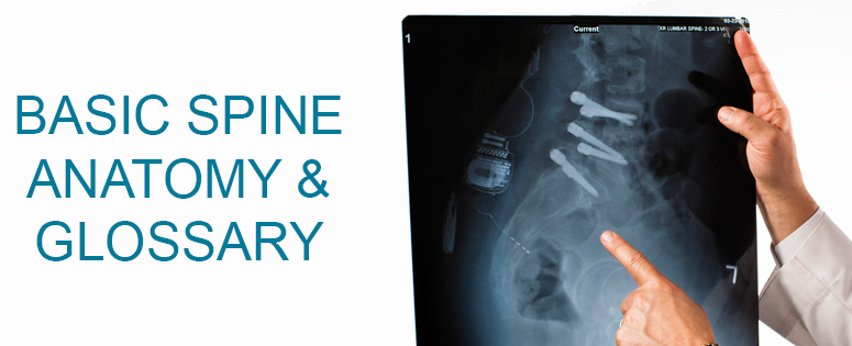
Understanding your spine and how it works together can help you understand some of the problems that occur from aging and/or injury. The spine is what allows you to hold up your head, shoulders and upper body. It allows you to stand up straight, bend and twist as well as protect the spinal cord.
Below is a glossary of the basic spine anatomy and conditions derived from spinal injury or disease. Tap or click on headings to show descriptions and definitions.
VERTEBRAE...The spine consists of small bones, called vertebrae (plural), which are stacked on top of another and together create the natural curves of the back.VERTEBRAE BODY...These bones that make up the spine create a canal or protective space for the spinal cord. The singular is vertebra.CERVICAL...The cervical spine is made up of 7 vertebrae that begin at the base of the skull.THORACIC...The thoracic spine is made up of 12 vertebrae that start in the upper chest and connect to the rib cage.
LUMBAR...The lumbar spine is made up of 5 vertebrae. These bones are larger than the cervical and thoracic vertebrae because they support more of the body’s weight.
SPINAL CORD...The spinal cords exits the skull and travels through each vertebral body in the center. This is called the spinal canal. At each level or at each vertebral body, there are nerve roots that exit and branch out to the muscles of the body. The openings where the nerve roots exit is called the neural formamin. The spinal cord typically ends at around the level of the first or second lumbar vertebrae. It then transforms into a bundle of nerve roots which is called the cauda equine.LAMINA...The lamina are portions of the bones on the back side of the spine that are arranged like shingles.FACET JOINT...The facet joint is the joint that connects each vertebral body to the next. This allows your spine to have movement and flexibility, including rotation. Just like the knee and hip joint, the facet joint can become arthritic and become a source of neck or back pain.DISC OR INTEVEBRAL DISC...The vertebral bodies of the spine are separated at each level with an intervertebral disc. They are flat, round and are about ½ inch thick. They consist of two things:
Nucleus pulposis which is a jelly-like substance that makes up the center of the disc. It contains water and this “cushion” allows the disk to be flexible.
Annulus fibrosis is the flexible and stronger outer ring of the disk. It has several levels similar to a rubber band.
Together, the nucleus pulposis and annulus fibrosis act as a “shock” absorber.COMPRESSION FRACTURE...A vertebral compression fracture occurs when the bones of the spine become broken due to trauma. Usually the trauma necessary to break the bones of the spine is quite large. In certain circumstances, however, such as in elderly people and in people with cancer, these same bones can break with little or no force. The vertebrae most commonly broken are those in the lower back.LAMINOTOMY...Is a procedure that removes part of a lamina of the vertebral arch in order to decompress the corresponding spinal cord and/or spinal nerve root. It increases the size of an opening in the lamina.LAMINECTOMY...This procedure involves removing the bony arch (lamina), any bone spurs, and ligaments that are compressing the spinal cord. Laminectomy relieves pressure on the spinal cord by providing extra space for it to drift backward.FUSION SPINAL FUSION...This is a surgical technique used to permanently join two or more vertebrae. Supplementary bone tissue, either from the patient (autograft) or a donor (allograft), is used in conjunction with the body's natural bone growth processes to fuse the vertebrae together. Fusing of the spine is used primarily to eliminate the pain caused by abnormal motion of the vertebrae by immobilizing the faulty vertebrae themselves, which is usually caused by degenerative conditions. However, spinal fusion is also the preferred way to treat most spinal deformities, specifically scoliosis and kyphosis.NON-UNION...A fusion that fails to heal means that there is no bone growth connecting the two levels. The two or more vertebral bodies fail to fuse.BONE GRAFTING...Bone grafting is a surgical procedure that replaces or augments missing bone in order to fuse the vertebral bodies together. Bone generally has the ability to regenerate but often it needs a “scaffold” to do so. Bone grafts may be autologous (bone harvested from the patient’s own body), allograft (cadaveric bone usually obtained from a bone bank), or synthetic (often made of hydroxyapatite or other naturally occurring and biocompatible substances) with similar mechanical properties to bone. Most bone grafts are expected to be reabsorbed and replaced as the natural bone heals over a few months’ time. BMPs (bone morphogenic proteins): BMPs are a group of growth factors that have an ability to induce the formation of bone and cartilage. BMPs can be synthetic and are often made of hydroxyapatite or other naturally occurring and biocompatible substances with similar mechanical properties to bone.EPIDURAL INJECTION...An epidural steroid injection is performed to help reduce the inflammation and pain associated with nerve root compression. Nerve roots can be compressed by a herniated disc, spinal stenosis, and bone spurs. When the nerve is compressed it becomes inflamed. The goal of the epidural steroid injection is to help lessen the inflammation of the nerve root.
The epidural space is located above the outer layer surrounding the spinal cord and nerve roots. An epidural steroid injection goes into the epidural space, directly over the compressed nerve root.
INTERLAMINAR AND TRANSFORMATIONAL INJECTIONS...They can also be described according to the path of the needle. Most epidural steroid injections are placed between the lamina, known as interlaminar epidural steroid injections. The lamina are portions of the bones on the back side of the spine that are arranged like shingles. The needle is aimed upwards toward the head and passes between two adjacent lamina. Another type of injection is a transforaminal steroid injection. In this case the needle passes along the course of the nerve and enters the spine from a more diagonal direction.
CAUDA EQUINA...In rare cases, a herniated disk may press on nerves that cause you to lose control of your bladder or bowel. If this happens, you may also have numbness or tingling in your groin or genital area. This is an emergency situation that requires surgery.SCIATICA...Sciatica is pain in your lower back or hip that radiates to the back of your thigh and into your leg. This may be due to a protruding (herniated) disk in your spinal column that is pressing on the roots of the sciatic nerve. This condition is known as sciatica.THECAL SAC...The thecal sac is the membrane that surrounds the spinal cord. The thecal sac is filled with cerebral spinal fluid.RADICULOPATHY...Nerve roots can be compressed by a herniated disc, spinal stenosis, and bone spurs. When the nerve is compressed it becomes inflamed. This can lead to pain, numbness, tingling or weakness along the course of the nerve. This is called radiculopathy.
SPINAL STENOSIS...The spinal cord travels through each vertebral body through the center. This is called the spinal canal. Spinal stenosis is when, due to various causes, the canal becomes narrowed and tight. There is less room for the spinal cord and this can cause pain in the back and legs. Patients with this problem often will report that bending or leaning forward while walking seems to relieve or lessen the pain.SCOLIOSIS...This is a type of spinal deformity that causes an abnormal curvature of the spine. When the spine is viewed from the front or back it should be in a straight line or like an “I”. Scoliosis is the sideways curve of the spine that when viewed from the back or front will look to an “S” or “C”.SPONDYLOLYSTHESIS...Spondylolysthesis: This often occurs as a result of degenerative disc disease and facet joint weakness. This is more commonly found in older black females and diabetics (affects females 6 times as much as males). This is when the vertebral body slips either forward or backwards with respect to the vertebral body below it.SPONDYLOSIS...This is a term referring to degenerative osteoarthritis of the joints between the centre of the spinal vertebrae and/or neural foraminae. If this condition occurs in the zygapophysial joints, it can be considered facet syndrome. If severe, it may cause pressure on nerve roots with subsequent sensory and/or motor disturbances, such as pain, paresthesia, or muscle weakness in the limbs.DEGENERATIVE DISC DISEASE (DDD)...This is not a disease but a term used to describe the normal changes in the discs as a person ages. These age related changes consist of loss of fluid in the discs. These discs will slowly over time “dry up” and the disc will narrow and be compressed. These changes are related to genetics, as well as if their job involves heavy manual labor. Smoking also predisposes patient’s to these changes. This most often occurs in the lumbar and cervical spine.HERNIATED DISC OR HERNIATED NUCLEUS PULPOSIS...Herniated Disc or Herniated Nucleus Pulposis: This is when the inside of a disc pushes outward or “herniates”. This often will compress a nerve root or even the spinal cord. Disc herniations can occur with or without any particular trauma.TORN ANNULUS...This is a tear or crack in the outer ring of the disc called the annulus fibrosis.OSTEOPHYTE...This is a bone growth or “bone spur” that often is a reaction by the body when there is degenerative disc disease. As the space between each vertebral body gets smaller there is less “padding” between them and there is more stress placed on the spine. Bone spurs can put pressure on nerves or the spinal cord.OSTEOPOROSIS...This is a disease of the bone that occurs when the creation of new bone doesn’t keep up with the removal of old bone. Bone is constantly regenerating and rebuilding. This most often happens in females and increases after menopause due to hormonal changes. People with osteoporosis are at risk of fracture.For more procedural information go to Treatments & Procedures

