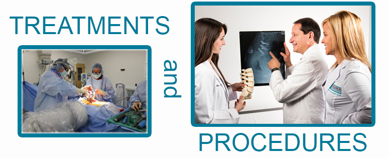
We try to begin with a conservative therapeutic approach, turning to surgery only when absolutely necessary. When surgery is the best option, we make all efforts to utilize minimally invasive techniques that enable patients to return to home, work and play as quickly as possible.
Below is a short glossary of procedures. For more information, get some video BACKTALK from Dr. Kirshner.
ACDF - ANTERIOR CERVICAL DISECTOMY AND FUSIONA small incision is made in the front of the neck to expose the front of the disc. The disc is removed to relieve pressure on the spinal cord and nerve roots. Once the herniated disc and/or bone spurs are removed, a cage device or bone graft is placed in between the vertebra to keep them from collapsing onto each other and to help them fuse or heal together. Then a small plate is placed over the vertebra and screws are used to attach the plate to the bones to keep them from moving so the fusion can heal.CADR - CERVICAL ARTIFICIAL DISC REPLACEMENTThe disc is removed from the front of the neck to relieve pressure from the spinal cord and nerve roots. Once this is completed, a disc prosthesis is inserted between the vertebral bodies. This keeps the bones separated as the normal disc would have and at the same time allows the bones to continue to move. This can lessen the mechanical stresses placed on the adjacent vertebra as may occur with a fusion.SPINAL FUSIONThis is a surgical procedure which is used to stabilize adjacent vertebra. This is done by roughening the bone surfaces and adding bone graft or bone graft extenders to help them heal together as if they were a single bone. Sometimes this will involve several vertebra in a row. This may also include some type of hardware (such as screws and plates or rods) which is used to help insure a successful healed fusion.MIS- MINIMALLY INVASIVE SURGERYThis is a technique not a procedure. MIS has evolved over the past several years, not just in spine surgery but in most of the surgical fields. We have found that the specific procedure, in many cases, can be performed through a smaller incision causing much less tissue damage. Typically when working on the lumbar spine, a decompression and fusion, including cages, pedicle screws and rods, can be done through two incisions (one on each side of the back) about 1 1/2 to 2 inches long. MIS surgery has been made possible primarily because of new technology. This includes magnification (microscope or magnification glasses), use of X-ray during the surgery and newer specially designed instruments.Studies have shown that this MIS technique is associated with:
- Significantly less blood loss, so transfusions are rarely needed
- Shorter hospital stays, most of our patients go home the same day or the next morning
- Less pain, the patient use less narcotic pain medication
- Patients are able to get back to work and their normal lives quicker.
POSTERIOR CERVICAL DECOMPRESSIONHere through an incision in the back of the neck part of the bone covering the back of the spinal cord and nerve roots is removed. This is done to relieve pressure which causes pain and weakness in the arms and sometimes the legs as well.
POSTERIOR CERVICAL FUSIONThis involves the placement of screws into the vertebra from the back of the spine then adding connecting rods to stabilize them. Once the hardware is in place bone graft is added to the roughened exposed boney surfaces so they will heal together as a single bone. This is often done in conjunction with a posterior decompression or an anterior cervical fusion.LUMBAR DISCECTOMYThis is the removal of a piece of the disc through a small incision in the lower back. After the 1 1/2 to 2 inch incision is made, the muscles are peeled away from a section of the bone. Next, small window of bone is removed to expose the nerves and the herniated disc. Now the piece of disc pinching and displacing the nerve is taken out. Typically this involves removal of less than 10% of the disc.ANNULAR REPAIROnce a disc herniates and the herniated portion is removed, there remains a hole in the disc. There is always a risk of the disc herniating again. When possible we try to identify that hole and close it with a device that is designed to lessen the risk of recurrent disc herniation.LUMBAR DECOMPRESSIONThis procedure is done through an incision in the lower back after gently pealing the muscles away from the bones covering the spinal canal. Once exposed the lamina and other compressing structures are gently removed until all of the pressure is relieved. This may involve one or several levels of the spine. This may include removing some of the bone, ligaments, discs and part of the facet joints.PLIF - POSTERIOR LUMBAR INTER-BODY FUSIONWorking from the back of the lumbar spine through one or several incisions, the disc is removed. Usually this includes removal of the lamina and one or both facet joints. This allows clear exposure of the disc and nerves. Next the disc is removed and the inner surfaces of the vertebra are roughened up to allow for bone healing. A cage and bone graft material is placed into the inter-body space. The cage device keeps the bones separated to prevent collapse and the bone graft material helps the bones to heal together. Next pedicle screws are place into the adjacent vertebra and metal rods are attached to the heads of the screws to stabilize the bones and prevent movement so they can heal. This is the same philosophy used to heal broken bones in other parts of the body such as the arm or leg. Once the hardware is in place, additional bone graft material may be added outside of the vertebra to further assist in the fusion and healing process.ALIF - ANTERIOR LUMBAR INTER-BODY FUSIONThis technique includes access to the lumbar spine through the belly. This is accomplished with the assistance of a general surgeon who helps to expose the front of the lumbar spine while protecting the abdominal organs and blood vessels. Once the exposure is completed, the entire disc is removed and the spinal canal is decompressed. Next a piece of bone or cage device filled with bone graft material is placed between the vertebra to keep them from collapsing on one another and to help them fuse together. The next step is to stabilize the spine which is done with a plate and screws in the front over the vertebra or from the back through an MIS or percutaneous technique.LADR - LUMBAR ARTIFICIAL DISC REPLACEMENTThis procedure involves the assistance of a general/vascular surgeon. This surgeon, through an incision in the belly, exposes the front of the lumbar spine protecting the abdominal and pelvic organs and blood vessels. Once the required region of the spine is exposed, we remove the damaged disc and relieve the pressure from the spinal canal and nerve roots. Now the artificial disc is placed between the exposed vertebra. There are metal endplates that are anchored to the end surfaces of the vertebra with a plastic spacer between them which allows for motion. This is similar in design to an artificial knee replacement.KYPHOPLASTYThis is a technique used to stabilize one or several vertebra that have been weakened by fracture and collapse. This can be caused by osteoporosis, trauma or tumor. The technique involves a small incision less than 1/4 of an inch. A small tube is placed into the broken bone through the pedicle from the back. Through this tube a balloon device is place into the fracture site and inflated to help restore the height of the bone and to make a void or an open hole in it. This hole is then filled with bone cement. This cement stabilizes the fracture therefore minimizing the pain and helping the patient get back to more normal function and mobility.XLIF - LATERAL LUMBAR INTER-BODY FUSIONThis technique involves an incision on the side. A tubular device is passed between the muscles onto the side of the disc. The soft tissues are cleared from that region and the tube is anchored to the adjacent vertebra. Using X-rays and direct vision, the disc is removed and the spinal canal decompressed. Once this is completed an inter-body cage is placed between the vertebra to keep them from collapsing and to assist in the fusion. The spine is then further stabilized with a lateral plate and screws through the same incision or with posterior pedicle screws and rods.SPINAL CORD STIMULATORThis procedure is done through the back. A small incision is placed in the mid-line of the middle back and a small laminotomy (window in the lamina) is made. Through this small opening a narrow electrode paddle is placed over the back of the spinal cord. This is then connected to a small stimulator pack which is placed in the lower back off to one side near the muscles. This is connected to the electrode paddle with a wire under the skin. This a procedure sometimes used to help minimize severe pain.More on Spine Anatomy/Glossary

